Stenting; NSCLC
56-year-old man was admitted to the department with complaints of continuous coughing and severe shortness of breath. CT of the chest showed 35-52mm tumor tissue, which partially blocked the lumen of the left main bronchus and completely blocked the lumen of the left lower lobe bronchus, causing atelectasis of the left lower lobe. Endobronchial biopsy showed adenosquamous carcinoma. Under general anesthesia 2 expandable, uncovered stents were inserted into the lower lobe and main bronchi. Bronchography taken after stent placement showed freely passed left main and lower lobe bronchus.
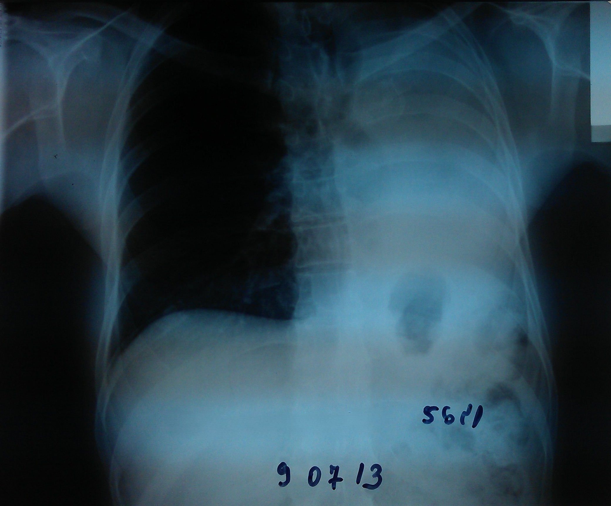
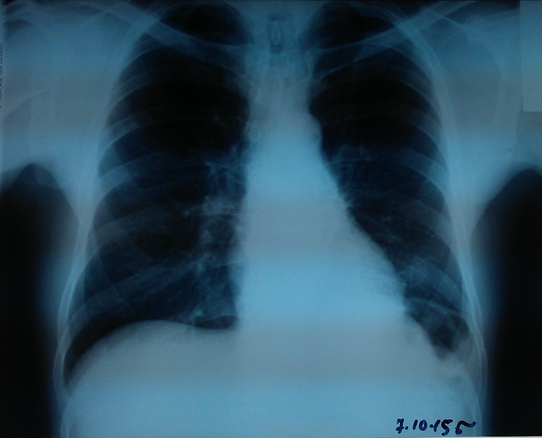
- Chest radiography 10 days prior to stent placement.
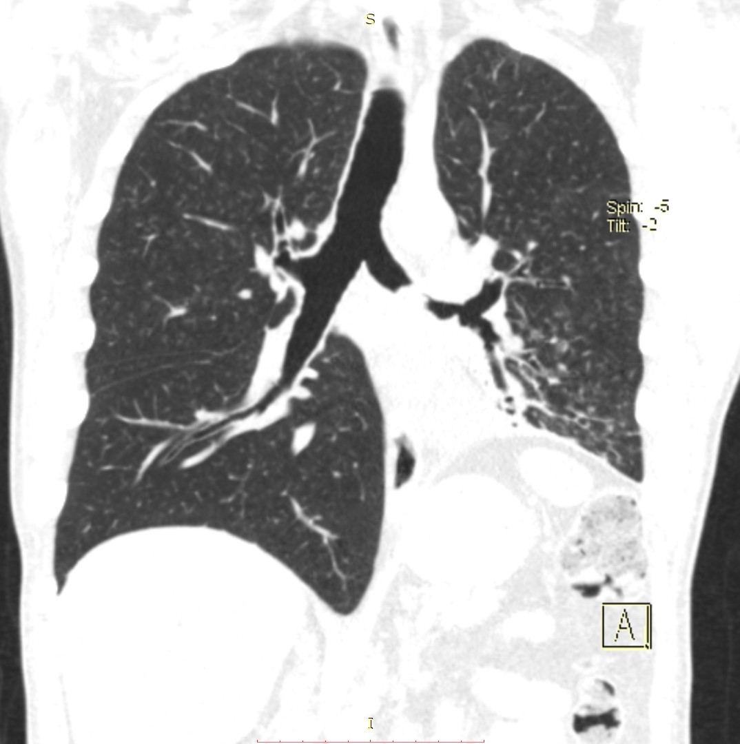
- Chest CT (coronal view) 5 days prior to stent placement.
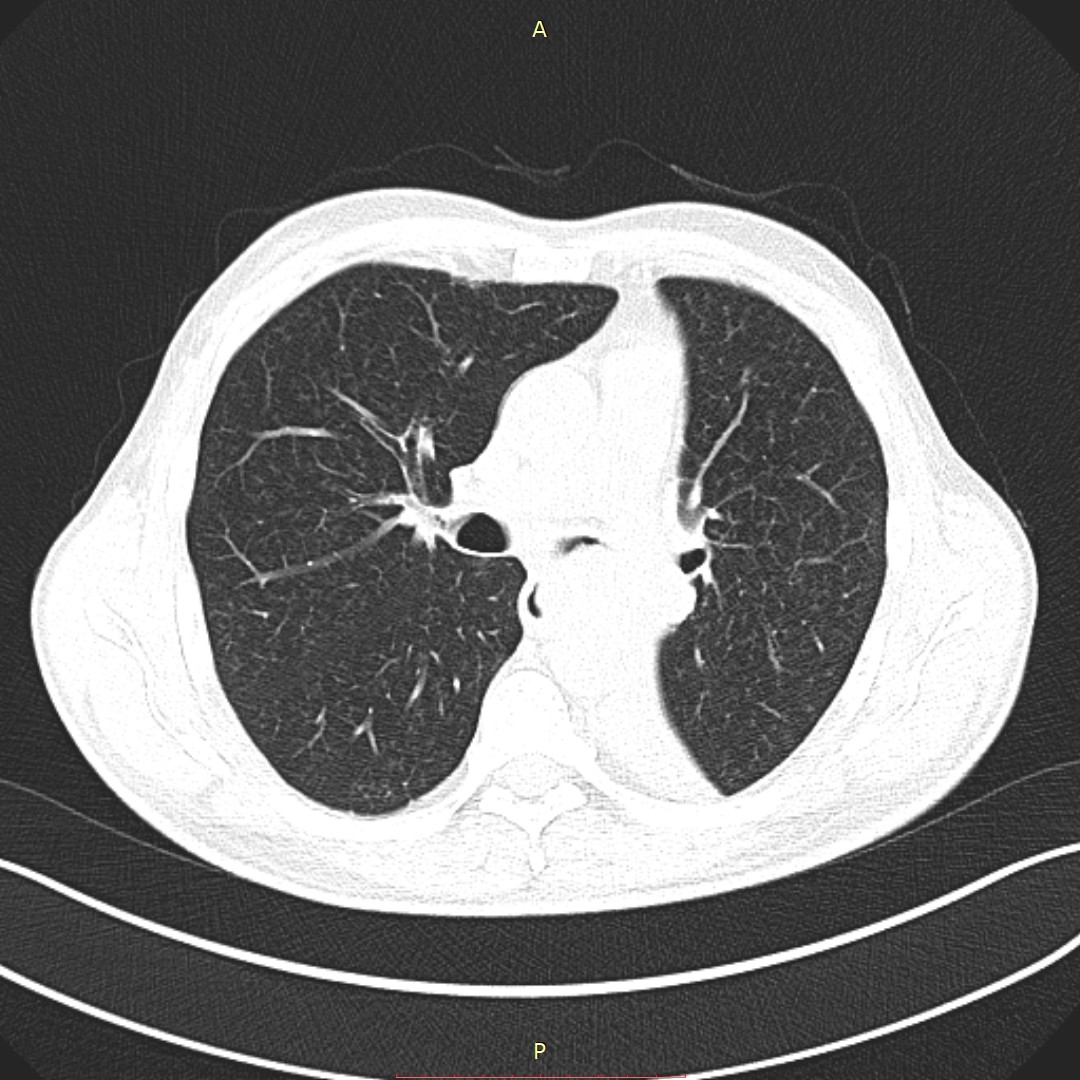
- Chest CT (axial view) 5 days prior to stent placement.
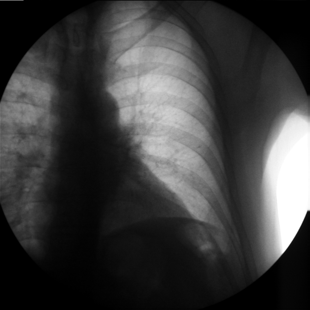
- Stenting of the left main and left lower lobe bronchi.
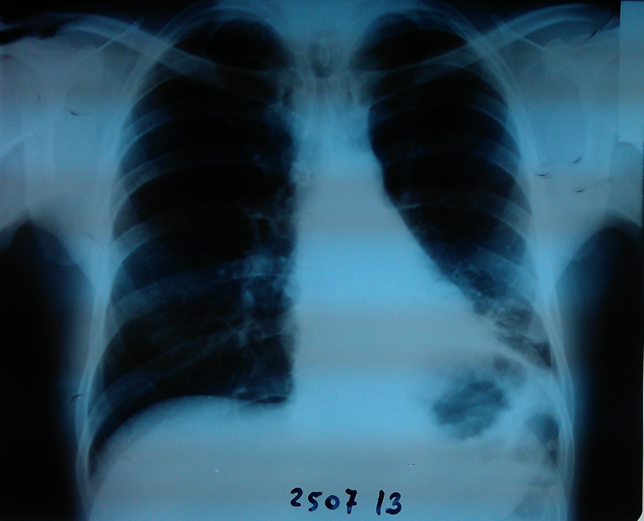
- Chest radiography 4 days after stent placement.
- Chest radiography 3 months after stent placement.
- Chest radiography 9 months after stent placement.