Bronchoscopic Lung Volume Reduction in Bullous Emphysema
A 42-year-old male patient, an active smoker, had been experiencing symptoms for several months.
Complaints: severe dyspnea on exertion, shortness of breath, and general weakness.
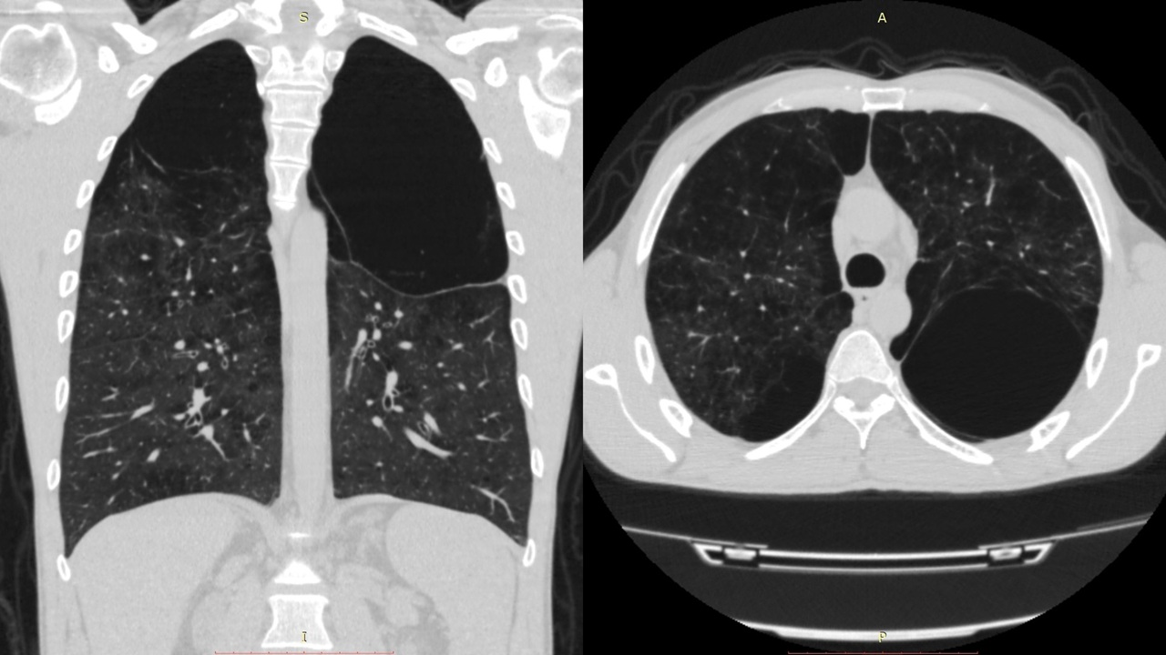
Chest CT findings (January 16, 2020): decreased lung parenchymal density in the upper lobes of both lungs, with giant bullae measuring 10–14 cm.
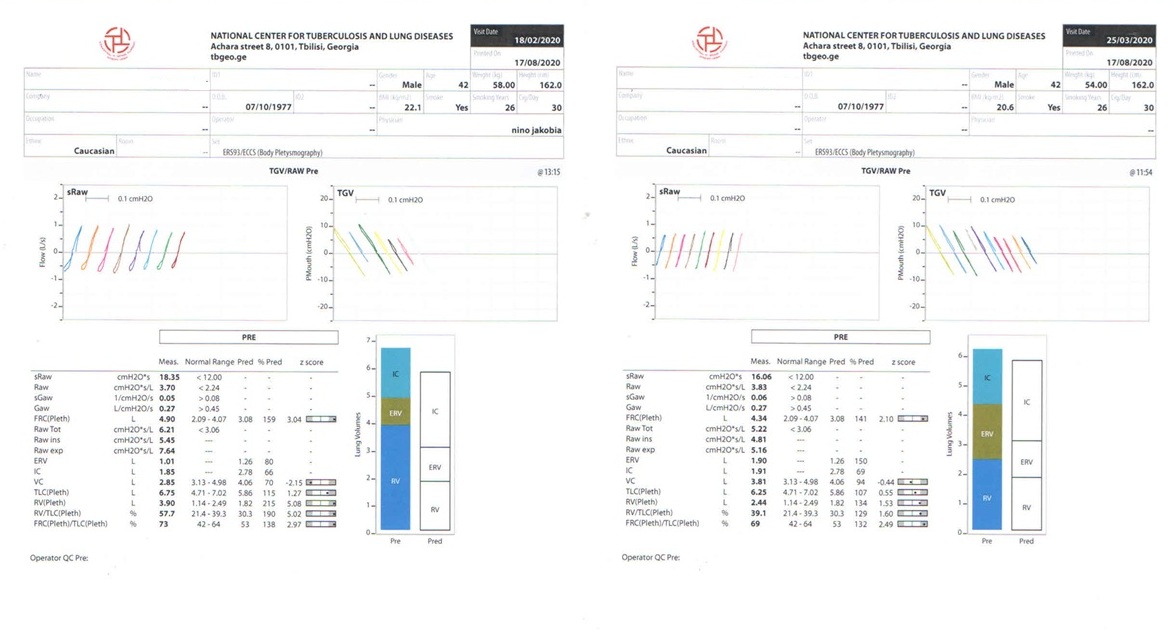
Body plethysmography (February 18, 2020):
RV (Pleth) – 3.90 L (normal range: 1.14–2.49 L).
On February 18, 2020, the patient underwent rigid bronchoscopy with valve bronchial occlusion for lung volume reduction of the left lung.
The procedure was performed under general anesthesia using a Friedel rigid bronchoscope, with a size 13 tube. A size 15 endobronchial valve (bronchial blocker) was placed in the lumen of the left upper lobe bronchus.
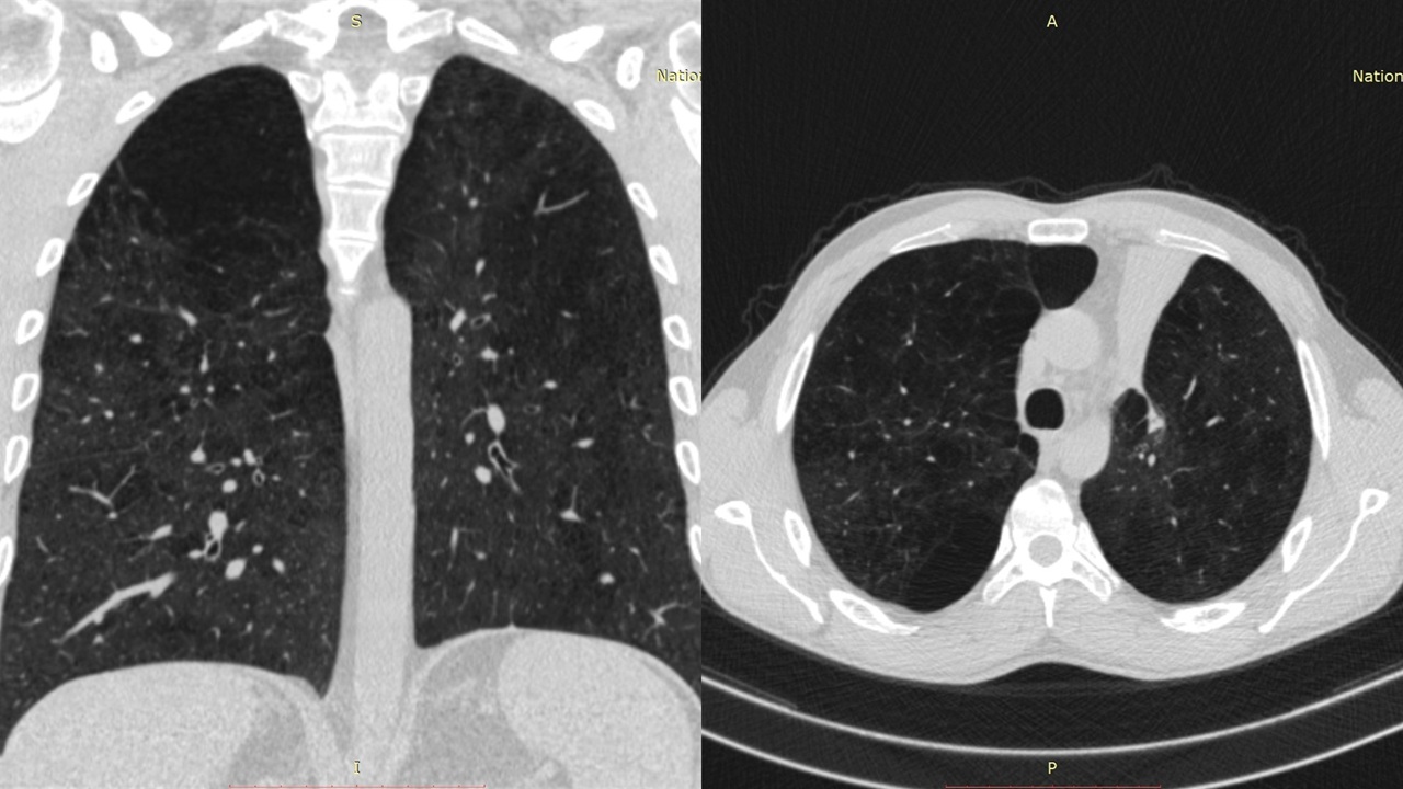
Follow-up at 3 months:
Chest CT demonstrated that the giant bulla in the left upper lobe was no longer visualized.
Body plethysmography (March 25, 2020):
RV (Pleth) – 2.44 L (normal range: 1.14–2.49 L).
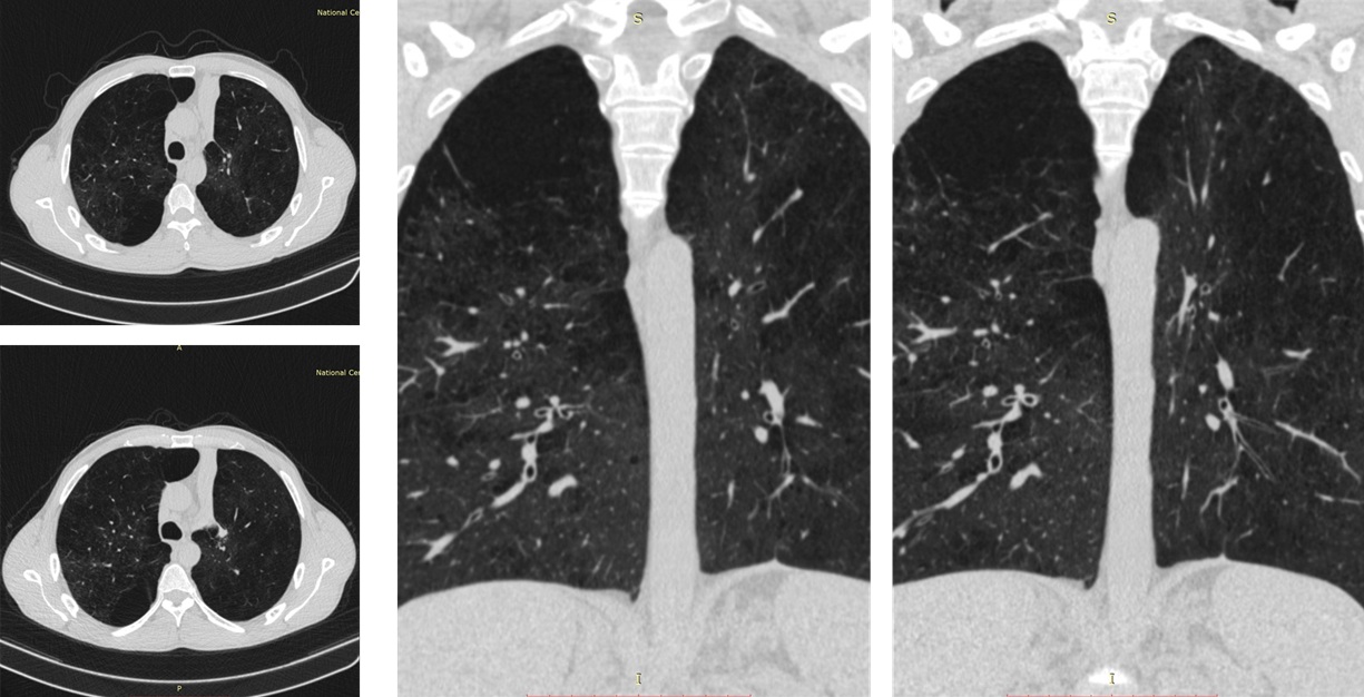
Positive radiological dynamics on chest CT were maintained at 9 and 12 months of follow-up.