Laser Resection; Mesenchymal origin Spindle Cell Tumour
56 year old male. Two months before admission the patient has dyspnea, spasmodic cough, appears fever.CT scan and bronchoscopy revealed endobronchial formation, which completely blocked the lumen of the left main bronchus. Endobronchial biopsy revealed Mesenchymal origin Spindle Cell Tumour. The patient’s condition worsened and under vital indications the laser resection was performed using VELASTM 30W Surgical Diode Laser System. After the recanalisation of the lumen from the lower lobe goes a lot of purulent mass. After antibiotic therapy the patient feels well.
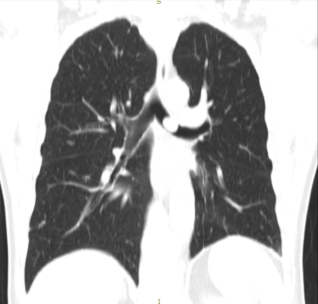
- Chest CT (coronal view) 2 weeks prior to resection.
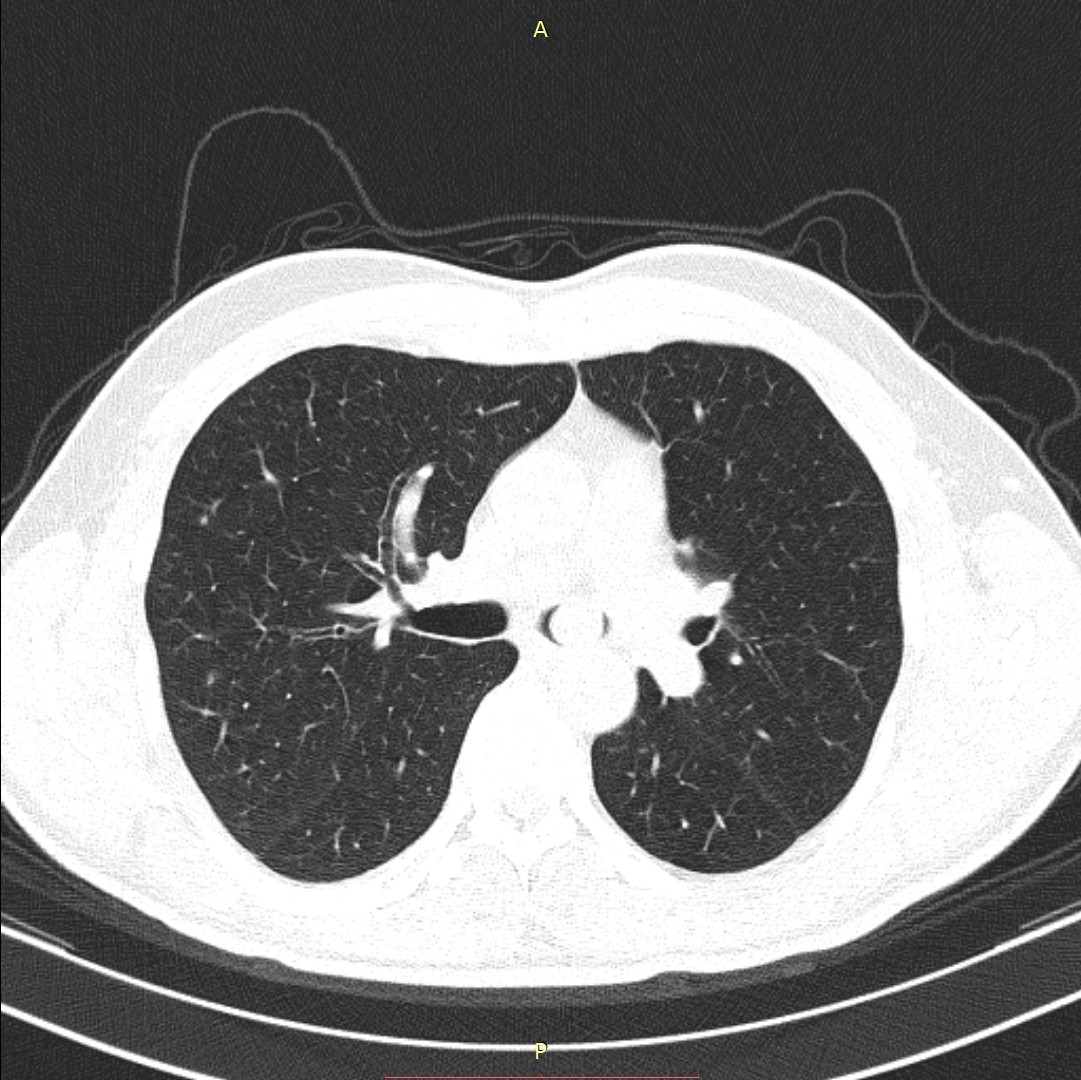
- Chest CT (axial view) 2 weeks prior to resection.
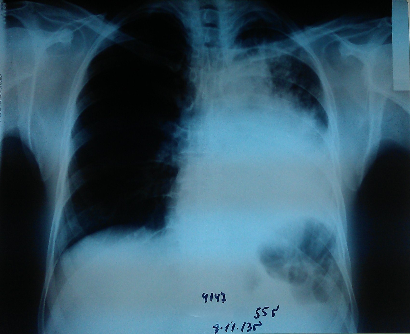
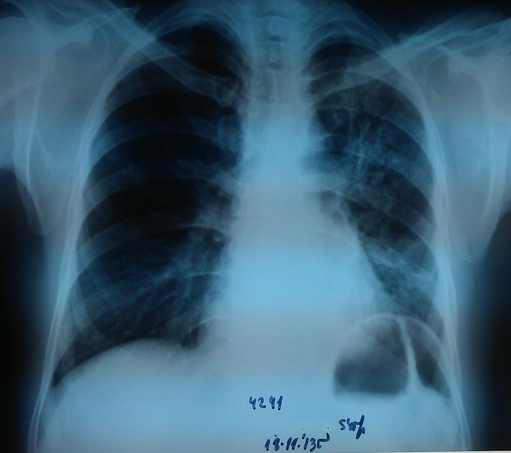
- Chest radiography 1 week prior to resection.
- Laser resection of the left main bronchus.
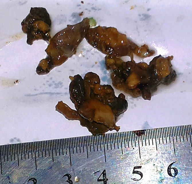
- Resected tumor fragments.
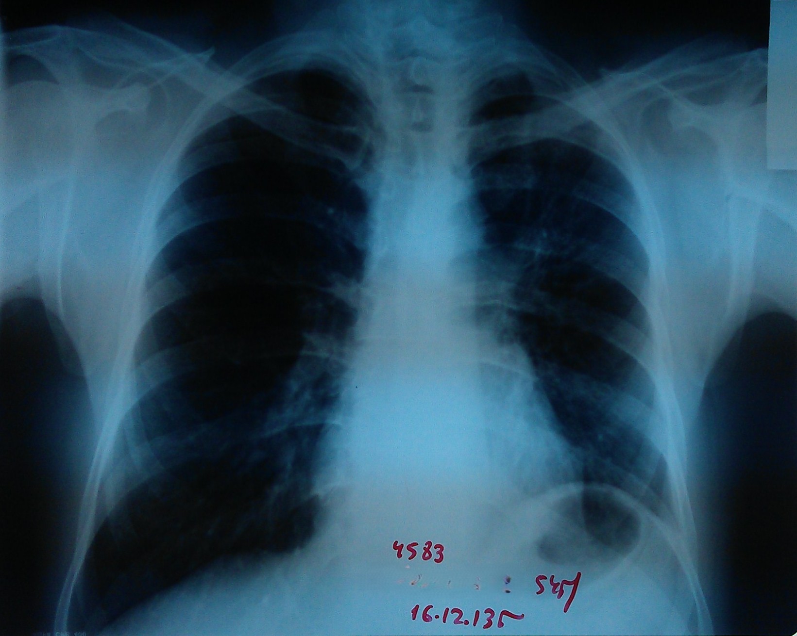
- Chest radiography 4 days after resection.
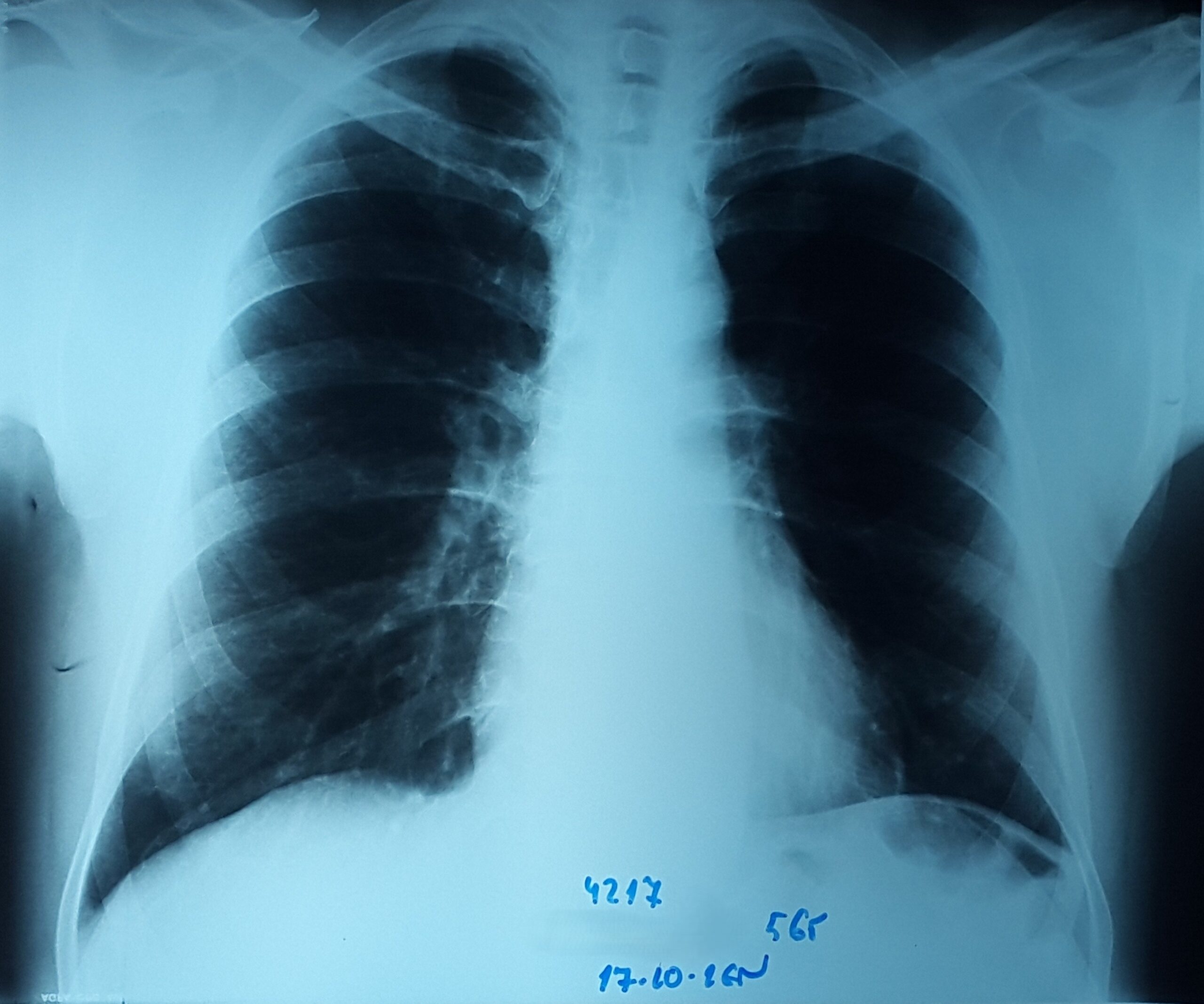
- Chest radiography 1 month after resection.
- Chest radiography 1 year after resection.
- Follow-up bronchoscopy 3 years after resection.